Abdominal Blood Vessels Labeled / Solved: Cadaver: Renal Blood Supply Label The Blood Vessel ... / Lateral view with the head to the right.. The videos are done by dr. Blood vessels of the abdomen and pelvis. The presence of vascular loops allows surgeons to ligate individual vessels with the expectation that blood will find its way to a particular region by alternate branches. Instant anatomy is a specialised web site for you to learn all about human anatomy of the body with diagrams, podcasts and revision questions Neurovasculature of the abdominal wall explore study unit superior epigastric artery:
Arteries are a type of blood vessel. Learn everything about the blood vessels of the abdominal wall with our article, quizzes, and labeled diagrams. The celiac trunk supplies blood to abdominal organs through three major subdivisions that branch from it, the common hepatic, left gastric, and splenic arteries. It is an artery, meaning that it carries blood away from the heart. Blood vessels of the abdomen and pelvis.
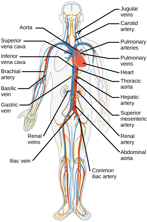
16.3: Circulatory and Respiratory Systems - Biology LibreTexts from bio.libretexts.org If you continue browsing the site, you agree to the use of cookies on this website. Superficial epigastric artery scheme of the femoral artery. They also take waste and carbon dioxide away from the tissues. The abdominal aorta enters the abdomen through the diaphragm at the level of the twelfth thoracic vertebre and continues to just below the umbilical area, where it splits into the right and left common iliac arteries. The common iliac arteries and veins. Nodes drain to preaortic lymph nodes in root of primary arteries of gut (celiac nodes, superior and iferior mesenteric nodes) Label the blood abdominal blood vessels labeled :. These vessels transport blood cells, nutrients, and oxygen to the tissues of the body.
They work to carry blood away from the heart.
Advertising on our site helps support our mission. They also take waste and carbon dioxide away from the tissues. Label the abdominal blood vessels using the hints provided. As the abdomen and pelvis contain the majority of internal organs, these regions need to be supplied by an extensive network of arteries and veins. Introductory anatomy lab #8 slideshare uses cookies to improve functionality and performance, and to provide you with relevant advertising. This video series covers the blood vessels for anatomy and physiology ii students. Development and function of the blood vessels: The common iliac arteries and veins. This study evaluates a cooperative learning approach for teaching anatomy to health science students incorporating small group and peer instruction based on the jigsaw method first described in the 1970's. Posterior cardinal sinus (blue) visible after lifting up the gi tract & gonads. These vessels are branches of the femoral artery and vein. It's the preferred screening method for an abdominal aortic aneurysm, a weakened, bulging spot in the abdominal aorta — the major blood vessel that supplies blood to the body. That being said, all arterial blood delivered to this region comes via branches of the abdominal aorta, and all venous blood eventually finds its way back to.
Development and function of the blood vessels: Lymph from abdominal viscera drains to lymphatic channesl and multiple named nodes alongisde arteries. These vessels are branches of the femoral artery and vein. (superficial epigastric vesseles labeled at center top.) details source femoral artery vein superficial epigastric vein identifiers latin arteria epigastrica superficialis mesh d019074 ta98 a12.2.16.011 ta2 4675 fma 20734 anatomical terminology [edit on. Instant anatomy is a specialised web site for you to learn all about human anatomy of the body with diagrams, podcasts and revision questions
Abdominal Blood Vessels from www.purposegames.com Instant anatomy is a specialised web site for you to learn all about human anatomy of the body with diagrams, podcasts and revision questions Blood vessels of the abdomen and pelvis. Label the blood abdominal blood vessels labeled :. Label the intestinal structures using the hints provided. However, the imaging test may be used to diagnose or rule out many other health conditions. (superficial epigastric vesseles labeled at center top.) details source femoral artery vein superficial epigastric vein identifiers latin arteria epigastrica superficialis mesh d019074 ta98 a12.2.16.011 ta2 4675 fma 20734 anatomical terminology [edit on. Within the thoracic abdominal region, the abdominal aorta gives rise to 10 major arterial branches that supply blood to organs, glands, bones, and other structures of the abdominal region. Want to learn more about it?
Doppler studies of the abdominal vessels demand an understanding of normal and abnormal blood flow patterns.
The videos are done by dr. Advertising on our site helps support our mission. Instant anatomy is a specialised web site for you to learn all about human anatomy of the body with diagrams, podcasts and revision questions Of course, recognition of the normal vascular anatomy is essential for the investigation of any abdominal vascular problem. Because arteries are moving blood being pumped out by the heart. Want to learn more about it? Doppler studies of the abdominal vessels demand an understanding of normal and abnormal blood flow patterns. That being said, all arterial blood delivered to this region comes via branches of the abdominal aorta, and all venous blood eventually finds its way back to. Learn everything about the blood vessels of the abdominal wall with our article, quizzes, and labeled diagrams. However, the imaging test may be used to diagnose or rule out many other health conditions. Lateral view with the head to the right. We will include an analysis of the normal doppler waveforms of the abdominal vessels. The venous drainage of the abdomen is carried out by the portal venous system and the systemic venous system.
Nodes drain to preaortic lymph nodes in root of primary arteries of gut (celiac nodes, superior and iferior mesenteric nodes) In contrast, veins carry blood back to the heart. Posterior cardinal sinus (blue) visible after lifting up the gi tract & gonads. (superficial epigastric visible at upper left.) the left femoral triangle. Aortas or aortae 4) is the main blood vessel in the abdominal cavity that transmits oxygenated blood from the thoracic cavity to the organs within the abdomen and to the lower limbs.
Vessel Lab from faculty.collin.edu Blood vessels of the abdomen and pelvis. The videos are done by dr. An abdominal ultrasound is done to view structures inside the abdomen. Aortas or aortae 4) is the main blood vessel in the abdominal cavity that transmits oxygenated blood from the thoracic cavity to the organs within the abdomen and to the lower limbs. If you continue browsing the site, you agree to the use of cookies on this website. Advertising on our site helps support our mission. Label the abdominal blood vessels using the hints provided. That being said, all arterial blood delivered to this region comes via branches of the abdominal aorta, and all venous blood eventually finds its way back to.
Label the blood abdominal blood vessels labeled :.
I hope this anatomy guide is helpful. Instant anatomy is a specialised web site for you to learn all about human anatomy of the body with diagrams, podcasts and revision questions The presence of vascular loops allows surgeons to ligate individual vessels with the expectation that blood will find its way to a particular region by alternate branches. An abdominal ultrasound is done to view structures inside the abdomen. Label the blood abdominal blood vessels labeled :. The aorta is the largest blood vessel in the body. The identification of abdominal vessels using ultrasound is based on knowledge of their normal location, appearance and relationship to specific organs. We will include an analysis of the normal doppler waveforms of the abdominal vessels. The celiac trunk supplies blood to abdominal organs through three major subdivisions that branch from it, the common hepatic, left gastric, and splenic arteries. Students were given time t … If you continue browsing the site, you agree to the use of cookies on this website. However, the imaging test may be used to diagnose or rule out many other health conditions. Label the biliary passages and associated structures using the hints provided.
If you continue browsing the site, you agree to the use of cookies on this website blood vessels labeled. Dissection of the blood vessels posterior to the diaphragm procedure:


Posting Komentar
0 Komentar